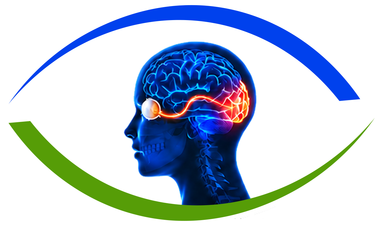
Visual field defects are blind spots or areas of diminished vision resulting from lesions along the anatomical visual pathway. Because the optic nerve fibers undergo specific decussation at the optic chiasm and project to the occipital visual cortex, the exact pattern of field loss (such as bitemporal hemianopia or homonymous hemianopia) provides localized neuro-anatomical mapping of tumors, strokes, or traumatic injuries.
Common Symptoms & Clinical Presentation
Patients with visual field defects commonly report one or more of the following characteristic symptoms:
- Bumping into doorframes, furniture, or pedestrians on one side of the body
- Difficulty reading lines of text or missing half of the printed page (hemianopic dyslexia)
- Impaired peripheral awareness while driving or crossing streets
- Loss of the outer temporal half of vision in both eyes (tunnel vision)
- Visual neglect or visual extinction in parietal lobe pathology
Underlying Causes & Mechanisms
Neuro-ophthalmic conditions frequently result from complex interactions between the central nervous system, autoimmune pathways, and vascular circulation:
- Chiasmal compression from pituitary adenomas, craniopharyngiomas, or meningiomas (bitemporal hemianopia)
- Ischemic stroke or intracranial hemorrhage in the posterior cerebral artery (PCA) territory
- Traumatic brain injury (TBI) affecting the temporal or parietal optic radiations
- Arteriovenous malformations (AVMs) or brain neoplasms
Specialized Diagnostic Workup
An accurate diagnosis requires in-depth tertiary evaluation that correlates visual sensory testing with computerized structural imaging:
- Standard Automated Perimetry (Humphrey Visual Field 24-2 and 30-2 testing)
- Goldmann kinetic visual field testing for peripheral field extent and legal driving qualification
- High-resolution Contrast MRI of the Brain, Sella Turcica, and Visual Pathways
- Spectral-Domain OCT with macular ganglion cell analysis (documenting retrograde trans-synaptic degeneration)
Management & Therapeutic Strategies
Treatment protocols are tailored to the specific etiology and severity of visual risk:
- Neurosurgical or endocrinological management of pituitary tumors (decompression/medical therapy)
- Visual field expansion prism therapy (Peli prisms) fitted to spectacles
- Specialized visual scanning and saccadic rehabilitation therapy
- Formal vision rehabilitation and occupational therapy for adaptive mobility
Frequently Asked Questions
Spontaneous neurological recovery occurs in about 30% to 50% of stroke patients within the first 3 months. Beyond 6 months, residual field loss is generally permanent, and focus shifts to compensatory scanning training and prism optics.
As a pituitary adenoma expands superiorly out of the sella turcica, it compresses the optic chiasm where nasal retinal fibers cross. This causes gradual, painless loss of the temporal (outer) visual field in both eyes (bitemporal hemianopia).